| The ureters deliver urine from the kidneys to the urinary bladder Click Here for Microscopic Image of Ureter |
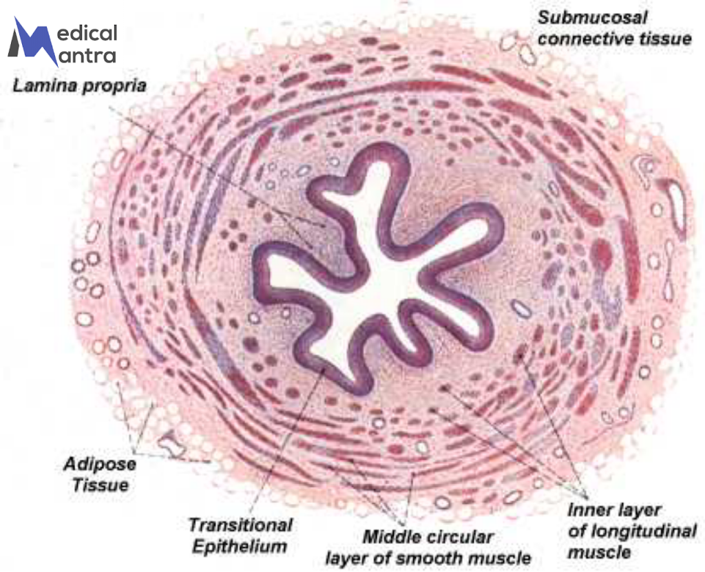
The ureter is a tube that connects the kidneys to the urinary bladder. The ureter is about 3 to 4 mm in diameter, and approximately 25 to 30 cm long . the ureter has three layers: the inner mucosa with folds, the middle muscularis with helically arranged muscle layers, and the outer fibrous connective tissue. Its main functions are to transport urine from the kidneys to the bladder using muscular contractions and to prevent urine from flowing back into the kidneys through muscular squeezing and flap-like structures at the ureter openings in the bladder.
Ureter has three main layers:
1. Mucosa: This is the innermost layer lining the ureter’s hollow space. It has folds that stick out into the tube when the ureter is empty. The mucosa is made up of a type of tissue called transitional epithelium, which is like a protective lining. It sits on top of a layer of connective tissue called the lamina propria.
2. Muscularis: This is the middle layer of the ureter and is responsible for the tube’s muscle contractions. It consists of two layers of smooth muscle cells. In the upper part of the ureter, the outer layer of muscle runs in circles, while the inner layer runs up and down. In the lower part of the ureter, an extra layer of muscle with fibers running up and down is added. These muscle layers are arranged in a helical pattern, giving the appearance of circular or up-and-down lines.
3. Fibrous Connective Tissue Covering: This is the outermost layer of the ureter and is made of fibrous connective tissue. It doesn’t have any special features and connects with the kidney’s outer covering at the top and with the bladder’s connective tissue at the bottom.
The ureter has two main functions:
1. Transporting Urine: The ureter’s job is to carry urine from the kidneys to the bladder. It does this by using muscular contractions that create waves, similar to how our intestines move food along through peristalsis. These contractions push the urine along the ureter and into the bladder.
2. Preventing Backflow: The muscular contractions of the ureter’s wall also help prevent urine from flowing back up into the kidneys. When the ureter enters the bladder, it passes through a muscular wall. This causes the ureter to get squeezed, preventing urine from flowing back. Additionally, there are flap-like structures made of mucosa (a protective layer) at the opening of each ureter in the bladder. These flaps act as valves, further preventing urine from flowing back into the ureters.


