Monday, October 27, 2025
  • About Us
  • Contact Us
  • Home
  • Privacy Policy
Medical Mantra
No Result
View All Result
  • Login
  • Register
  • News
  • Notes
    • All
    • Anatomy
    • gentics
    • Pathology
    Classification of NEOPLASIA

    Classification of NEOPLASIA

    Thalassemia

    Thalassemia: Types, Symptoms, Causes and Treatment

    Techniques for the Study of Pathology

    Techniques for the Study of Pathology

    Pathology Overview : Definition, Methods, Concepts

    Pathology Overview : Definition, Methods, Concepts

    Introduction of the Anatomy of the Upper Limb: Evolutionary Adaptations and Functional Complexity

    Introduction of the Anatomy of the Upper Limb: Evolutionary Adaptations and Functional Complexity

    Glomerulonephritis : Types, Causes, Symptoms, Diagnosis and Treatment

    Glomerulonephritis : Types, Causes, Symptoms, Diagnosis and Treatment

    Trabeculae Bone: Anatomy, Structure, Function,

    Trabeculae Bone: Anatomy, Structure, Function,

    Congenital Malformation: An Overview of Causes, Mechanisms, and Treatment

    Congenital Malformation: An Overview of Causes, Mechanisms, and Treatment

    Trisomy X Syndrome (47,XXX) : Causes, Symptoms, Diagnosis, Treatment

    • Anatomy
    • Pathology
    • gentics
  • Quiz
    Quiz Template Cell Injury

    Cell injury Quiz – Pathophysiology

  • Image Bank
  • Clinical Cases
No Result
View All Result
  • Login
  • Register
Medical Mantra
No Result
View All Result
Home histology slides

Spleen Histology : General Overview, Structure, Vascular Supply , Regions and Layers , Function of Spleen

Medicalmantra.org by Medicalmantra.org
May 29, 2025
in histology slides, Image Bank
0
Spleen Histology : General Overview, Structure, Vascular Supply , Regions and Layers , Function of Spleen
0
SHARES
75
VIEWS
Share on FacebookShare on Twitter

Spleen Histology : General Overview, Structure, Vascular Supply , Regions and Layers , Function of Spleen
The spleen, the largest lymphoid organ in the body, is
invested by a collagenous connective tissue capsule; it
has a convex surface and a concave aspect, known as
the hilum.
Click Here For More Microscopic Images


General Information of Spleen:

  • The spleen is the largest lymphoid organ in the body.
  • It weighs approximately 150 g in an adult individual.
  • It is located in the upper left quadrant of the abdominal cavity, within the peritoneum.
  • The spleen functions in immune responses, blood filtration, and the destruction of old red blood cells and platelets.
  • During fetal development, the spleen is involved in the production of blood cells.

Structure of Spleen:

  • The spleen is surrounded by a dense, irregular fibroelastic connective tissue capsule.
  • The capsule is covered by visceral peritoneum, which provides a smooth surface for the spleen.
  • The spleen has a convex surface and a concave aspect known as the hilum.
  • The hilum is the site where arteries, veins, and lymph vessels enter and leave the spleen.
  • Trabeculae, arising from the capsule, carry blood vessels into and out of the spleen.
  • The spleen has a three-dimensional network of reticular fibers and reticular cells, which form its architectural framework.
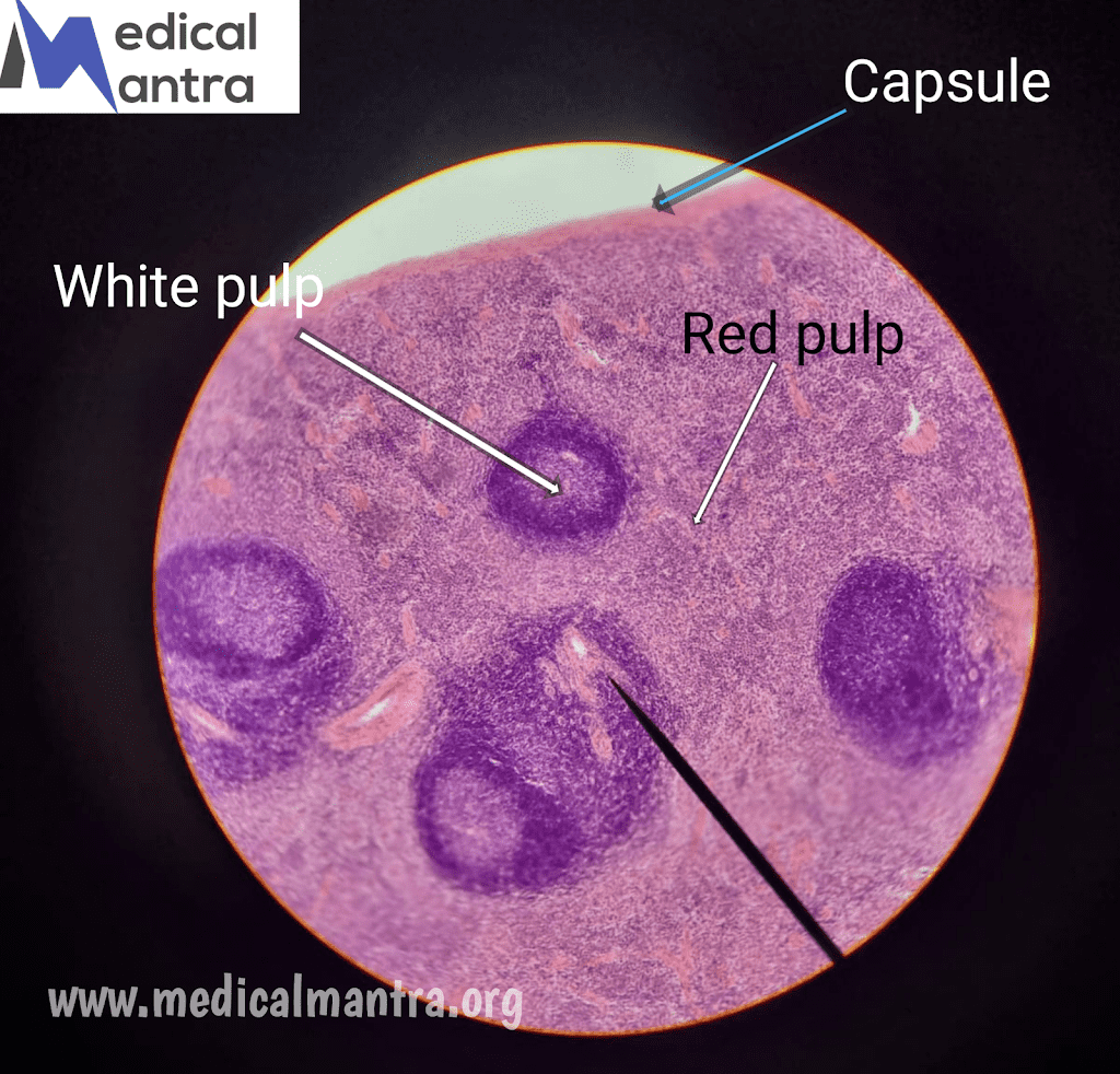
  • The splenic parenchyma consists of white pulp and red pulp.
  • White pulp is mainly composed of lymphocytes, while red pulp contains splenic sinuses and splenic cords.

Regions and Layers of Spleen:

White pulp: 

  • It is composed of periarterial lymphatic sheath (PALS) and lymphoid nodules.
  • PALS surrounds the central artery and contains T lymphocytes.
  • Lymphoid nodules are composed of B cells and can have germinal centers.
  • Marginal zone: It separates the white pulp from the red pulp and contains plasma cells, T and B lymphocytes, macrophages, and dendritic cells.

Red Pulp:

  • Red pulp consists of splenic sinuses and splenic cords.
  • Splenic sinuses have fusiform endothelial cells with spaces between them.
  • Reticular fibers surround the sinuses, and macrophages are numerous in their vicinity.
  • Splenic cords are a loose network of reticular fibers with extravasated blood.
  • Macrophages in the red pulp phagocytose blood-borne antigens, bacteria, and old platelets and erythrocytes.
Blood Supply and Circulation of Spleen:
  • Vascular Supply: The spleen is supplied by the splenic artery and drained by the splenic vein, both entering and leaving the spleen at the hilum.
  • Trabecular Arteries: Branches of the splenic artery, called trabecular arteries, penetrate the capsule and carry blood into the spleen.
  • Central Artery: The central artery, located in the periarterial lymphatic sheath, gives rise to follicular arterioles, supplying the lymphoid nodules.
  • Red Pulp Circulation: From the central artery, blood flows into the red pulp through penciller arterioles. Blood is then drained into splenic sinuses and eventually leaves the spleen through the splenic vein

Functions of Spleen:

Immunological Functions:

  • Antibody Formation: The spleen plays a role in the production of antibodies by B cells in the lymphoid nodules.
  • T-Cell and B-Cell Proliferation: The white pulp of the spleen supports the proliferation of T cells and B cells in response to antigens.
  • Immune Response Activation: The spleen acts as a site for immune responses, where lymphocytes interact with antigens presented by dendritic cells.

Blood Filtration and Clearance:

  • Blood Filter: The spleen filters the blood, removing debris, microorganisms, and blood-borne antigens.
  • Removal of Old Blood Cells: Macrophages in the spleen phagocytose and remove aged red blood cells and platelets.
  • Clearance of Damaged Cells: The spleen plays a role in the clearance of damaged or defective platelets and neutrophils.

Hemopoiesis:

  • Fetal Hemopoiesis: During fetal development, the spleen is involved in the production of blood cells.
  • Adult Hemopoiesis: In adults, hemopoiesis primarily occurs in the bone marrow, but the spleen can resume its hemopoietic function if needed.

Tags: histological slideshistologyspleen histology

RelatedPosts

Unlock Tonsils Histology:  Learn the Structure and Composition of Palatine Tonsils, Pharyngeal Tonsils, Lingual Tonsils
histology slides

Unlock Tonsils Histology: Learn the Structure and Composition of Palatine Tonsils, Pharyngeal Tonsils, Lingual Tonsils

by Medicalmantra.org
May 29, 2025
Lymph Nodes Histology: Learn the Structure, Type, Zone, and Functions of Lymph Nodes
histology slides

Lymph Nodes Histology: Learn the Structure, Type, Zone, and Functions of Lymph Nodes

by Medicalmantra.org
May 29, 2025
Red Bone Marrow Histology: Zones, Cells, and Functions of Red Bone Marrow
histology slides

Red Bone Marrow Histology: Zones, Cells, and Functions of Red Bone Marrow

by Medicalmantra.org
May 29, 2025
Thymus Histology : General Overview, Structure, Layers, Vascular Supply, and Functions of The Thymus.
histology slides

Thymus Histology : General Overview, Structure, Layers, Vascular Supply, and Functions of The Thymus.

by Medicalmantra.org
May 29, 2025

Leave a Reply Cancel reply

Your email address will not be published. Required fields are marked *

Medical Mantra logo

Medical Mantra is a dedicated platform that provides high-quality, evidence-based content for medical students and doctors. Our mission is to make medical education simple, accessible, and engaging — through clinical case studies, trending articles, quizzes, and community discussions. With Medical Mantra, learning is not just a requirement, it's a journey.

Pages

  • About Us
  • Contact Us
  • Home
  • Privacy Policy

Quick links

  • About Us
  • Contact Us
  • Privacy Policy
No Result
View All Result

Categories

Archives

  • About Us
  • Contact Us
  • Home
  • Privacy Policy

© 2025 Copyright Medicalmantra.org - Created and Designed by Medical Mantra.

Welcome Back!

Sign In with Google
Sign In with Linked In
OR

Login to your account below

Forgotten Password? Sign Up

Create New Account!

Sign Up with Google
Sign Up with Linked In
OR

Fill the forms below to register

All fields are required. Log In

Retrieve your password

Please enter your username or email address to reset your password.

Log In
No Result
View All Result
  • Home
  • Blogs
  • Contact Us
  • Privacy Policy
  • About Us

© 2025 Copyright Medicalmantra.org - Created and Designed by Medical Mantra.